


PRODUCTS BY SECTOR


Sacrum Ultrasound Scanning – Measuring Effectiveness
For optimum pressure relief under boney prominences Therawave mattresses utilise dedicated pressure area zones that provide individualised therapy underneath high risk areas such as the heels and sacrum. The levels of oedema within dermal tissue on these areas can be quantitatively assessed through the use of high definition ultrasound analysis.
Each ultrasound scan of the tissue is analysed using a form of pixel distribution analysis whereby pixels below certain intensity are classed as Low Echogenic Pixels (LEP). The ratio of LEP’s to Total Pixel count (TP) has been shown to reflect changes in dermal water content
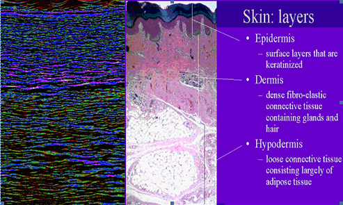
Below is a scan of normal skin with adjacent histology section indicating zones.
Below are ultrasound scans that show the performance of a Therawave 8 alternating mattress on a paitent with sacrum wound damage over a 4 week period.
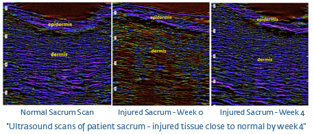
It can be seen from the scans that the normal skin dermis is mainly composed of fibrous tissue which ultrasonically is shown as mainly blue pixels. Compare this to the injured tissue at time 0 weeks which has a large number of red pixels in the scan. This is a typical profile of an oedematous tissue. The scan of the same tissue after 4 weeks of the patient on a Therawave mattress shows that there is a decrease in red pixels and an increase in blue pixels which indicates a movement back to the uninjured state.
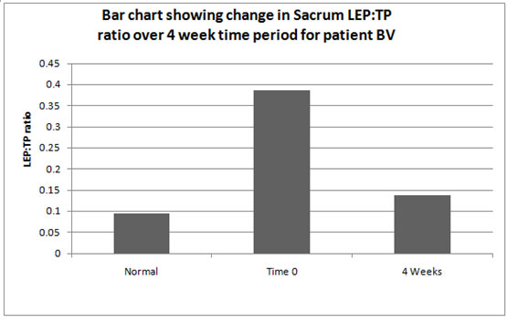
The graph above shows that the LEP:TP ratio after initially moving away from normal uninjured levels returns back to normal levels within 4 weeks after starting therapy on a Therawave mattress.